KPV (Lys-Pro-Val) produces reproducible anti-inflammatory effects in cell and mouse colitis models through PepT1-mediated uptake and NF-κB inhibition. The evidence is consistent across at least three independent research groups working in intestinal, dermatological, and oncological contexts. What it does not have is a single completed human randomized controlled trial, and it carries no FDA approval for any indication. Every legitimate supply channel treats it as a research-use-only compound.
TL;DR:
- KPV’s efficacy depends heavily on PepT1 expression, which must be confirmed in the target tissue before expecting therapeutic effects.
- The lipophilic nanoparticle formulation with hyaluronic acid and hydrogel improves tissue targeting and stability, enhancing delivery over free KPV.
- No human safety data exists, and all current studies are limited to mice and cell models, so clinical application remains unproven.
- Consistent, detailed analysis of peptide purity and formulation parameters is essential to ensure experimental reproducibility and interpretability.
- Future research should focus on pharmacokinetic studies in larger animals and human tissue profiling to validate PepT1 dependence and dosing strategies.
Table of Contents
- What Is KPV Peptide Research Actually Studying?
- Key Preclinical Studies Behind KPV's Anti-Inflammatory Profile
- Nanoparticle and Hydrogel Delivery: Why Formulation Changes the Outcome
- Safety Data, Sourcing Risk, and Regulatory Status
- Where KPV Research Needs to Go Next
- Verifying Compound Quality for KPV Protocols
- The Gap Between a Good Mechanism and a Proven Treatment
- Sources
- FAQ
What Is KPV Peptide Research Actually Studying?
KPV is the C-terminal tripeptide fragment of alpha-melanocyte-stimulating hormone (α-MSH), stripped down to three amino acids: lysine, proline, and valine. That truncation matters more than it sounds. Full-length α-MSH signals through melanocortin receptors, which also drive pigmentation and a cascade of endocrine effects that complicate any therapeutic use. KPV does not activate those receptors at all, yet it retains the anti-inflammatory punch of its parent molecule. Researchers investigating KPV are essentially asking one question: can you isolate the immune-modulating piece of α-MSH biology without dragging along the melanocortin baggage?
The answer, at least in preclinical models, looks like yes. The transporter that makes this possible is PepT1 (SLC15A1), a proton-coupled oligopeptide transporter normally responsible for absorbing dietary di- and tripeptides in the small intestine. Under inflammatory conditions, PepT1 expression climbs in colonic epithelium, a shift documented in both animal models of colitis and human biopsy tissue. That upregulation happens to occur precisely where researchers want a therapeutic peptide to concentrate. KPV rides that transporter directly into inflamed tissue.
Once inside the cell, KPV does not sit idle. It interferes with the nuclear factor kappa B (NF-κB) and MAPK signaling cascades that drive transcription of pro-inflammatory cytokines. This is the mechanistic core of nearly every KPV study published so far, and it explains why the peptide shows activity in tissues as different as gut epithelium and skin keratinocytes despite sharing no obvious anatomical similarity.
A few structural and kinetic details define how this plays out at the molecular level:
- KPV binds hPepT1 with a low Km in intestinal cell lines, meaning meaningful intracellular accumulation happens at low extracellular concentrations rather than requiring pharmacologic excess.
- Genetic knockout or pharmacological silencing of PepT1 abolishes KPV's anti-inflammatory effect in mouse models, confirming the transporter is not incidental but required.
- Downstream, KPV suppresses IκB degradation, which keeps NF-κB sequestered in the cytoplasm and blunts transcription of TNF-α, IL-6, and IL-1β.
- The peptide shows no measurable affinity for melanocortin receptors 1 through 5, separating its anti-inflammatory action from any pigmentation or appetite-related endocrine signaling.
Statistic Callout: PepT1 dependence is not a minor caveat. In PepT1-knockout mice, KPV's protective effects against colitis and colitis-associated tumor formation disappear entirely, according to work extending the original transporter research. That single finding is why any translational program has to start with confirming PepT1 expression in the target tissue before assuming the peptide will do anything at all.
The kinetic profile also explains something researchers often ask about early: why a tripeptide, rather than a larger peptide or small molecule, works here. Larger peptides generally do not have a dedicated intestinal transporter waiting to usher them into cells. KPV's three-residue length happens to fit PepT1's substrate pocket, and that structural coincidence is arguably the single most important fact in the entire body of KPV research. Without it, the peptide would likely degrade in the gut lumen before reaching any target.
Key Preclinical Studies Behind KPV's Anti-Inflammatory Profile
The foundational study in this field came from Dalmasso and colleagues, published in Gastroenterology. Their group established that orally administered KPV, delivered in drinking water at approximately 205 micrograms per day, reduced disease severity in both dextran sulfate sodium (DSS) and 2,4,6-trinitrobenzene sulfonic acid (TNBS) induced colitis models in mice. These two models trigger inflammation through different mechanisms (DSS damages epithelial barrier integrity directly, while TNBS provokes a T-cell mediated immune response), so consistent efficacy across both suggests the anti-inflammatory action is not model-specific.
Endpoints in that study included body weight change, colon length (shortening is a reliable proxy for inflammatory severity in mice), myeloperoxidase (MPO) activity as a marker of neutrophil infiltration, histological scoring of tissue damage, and direct measurement of pro-inflammatory cytokine expression. Treated mice showed reduced colonic shortening, lower MPO activity, and blunted cytokine expression relative to untreated colitis controls. Critically, the same paper demonstrated that KPV is directly transported into intestinal epithelial and immune cells via hPepT1, tying the functional outcome back to the mechanistic story.
Subsequent work by Viennois and colleagues extended this line of research into colitis-associated cancer, a clinically relevant progression since chronic ulcerative colitis substantially raises colorectal cancer risk. Their experiments reinforced the PepT1-dependence finding: knock out the transporter, and KPV's protective effects against both colitis and downstream tumor formation vanish. This second, independent confirmation of the PepT1 requirement is part of what makes the mechanism credible rather than a single-lab artifact.
A more recent line of investigation moved outside the gut entirely. A study on KPV and keratinocyte oxidative stress examined HaCaT keratinocytes exposed to PM10 particulate matter, a common model for pollution-induced skin damage. KPV reduced reactive oxygen species (ROS) accumulation and IL-1β secretion, and it attenuated PM10-induced cytotoxicity, with effects also demonstrated in a 3D skin model. The signaling pathway implicated, MAPK/NF-κB modulation, overlaps substantially with the gut mechanism, which is a meaningful cross-validation. A peptide hitting the same pathway in two unrelated tissue types is a stronger signal than either finding alone.
| Study | Model | Route / Dose | Key Endpoints | Primary Finding |
|---|---|---|---|---|
| Dalmasso et al. | DSS and TNBS mouse colitis | Oral, ~205 µg/day in drinking water | Weight, colon length, MPO, histology, cytokines | Reduced inflammation via PepT1-mediated uptake and NF-κB inhibition |
| Viennois et al. (extension) | DSS colitis and colitis-associated cancer, PepT1-KO mice | Oral administration | Tumor incidence, colitis severity | Efficacy is fully PepT1-dependent; lost in knockout mice |
| Keratinocyte PM10 study | HaCaT cells, 3D skin model | In vitro exposure | ROS levels, IL-1β secretion, viability | Reduced oxidative stress and cytokine release via MAPK/NF-κB |
Several limitations run through all three lines of work. Mouse colitis models, while standardized, do not fully replicate the chronicity or genetic heterogeneity of human inflammatory bowel disease. Pharmacokinetic and pharmacodynamic parameters in larger mammals remain uncharacterized. And oral bioavailability in mice, where gut transit time and pH profile differ substantially from humans, may not predict human absorption at all.
Researchers designing follow-up work should build in a few standard controls that not every early study included consistently:
- A PepT1 inhibitor or knockout arm to confirm transporter dependence in the specific model being tested.
- Vehicle-only and disease-model-only controls run in parallel, not historical comparisons.
- Dose-response arms rather than a single fixed dose, since the 205 µg/day figure from the original mouse study has rarely been systematically varied.
- Blinded histological scoring to reduce observer bias in tissue damage assessment.
Nanoparticle and Hydrogel Delivery: Why Formulation Changes the Outcome
Free KPV faces a practical problem before it ever reaches PepT1: the gastrointestinal tract is full of proteases eager to degrade small peptides before they cross the epithelium. Formulation is often the rate-limiting factor between a peptide that works in a dish and one that works when swallowed, and KPV research illustrates this clearly.
The most developed solution so far uses hyaluronic acid-functionalized PLGA nanoparticles loaded with KPV and then embedded in a chitosan/alginate hydrogel. The rationale has two layers. First, PLGA (poly lactic-co-glycolic acid) is a biodegradable polymer that shields the peptide payload from enzymatic breakdown during transit. Second, hyaluronic acid functionalization gives the nanoparticle affinity for CD44, a cell-surface receptor overexpressed on inflamed and activated immune cells in the gut, adding a second layer of targeting on top of PepT1 uptake itself. The chitosan/alginate hydrogel shell adds a third mechanism: pH-triggered release timed to survive the stomach's acidity and disintegrate closer to the colon, where the target tissue actually sits.
In the reported formulation, the HA-KPV nanoparticles measured a size favorable for cellular uptake with a negative zeta potential, a size range generally favorable for cellular uptake without triggering rapid clearance. In a DSS-induced mouse colitis model, this formulation improved colonic targeting, accelerated mucosal healing, and reduced inflammatory markers compared with non-functionalized nanoparticle controls. Treated tissue histology looked comparable to healthy controls in several measures, a result that free, unprotected KPV did not match at equivalent doses.
Pro Tip: If you're evaluating a KPV formulation for a preclinical protocol, don't take particle size or zeta potential figures from a supplier data sheet at face value. Ask for the actual release kinetics profile under simulated gastric and colonic pH, plus a cell uptake assay in a relevant line, since these determine whether the peptide reaches its target intact far more than the raw size number does.
That gap between free and encapsulated KPV performance is arguably the most practically important finding in this entire body of research for anyone planning translational work. A researcher who only reads the Dalmasso mouse data might reasonably expect an oral KPV protocol to work as-is. The nanoparticle work suggests otherwise, at least for reaching therapeutic concentrations efficiently.
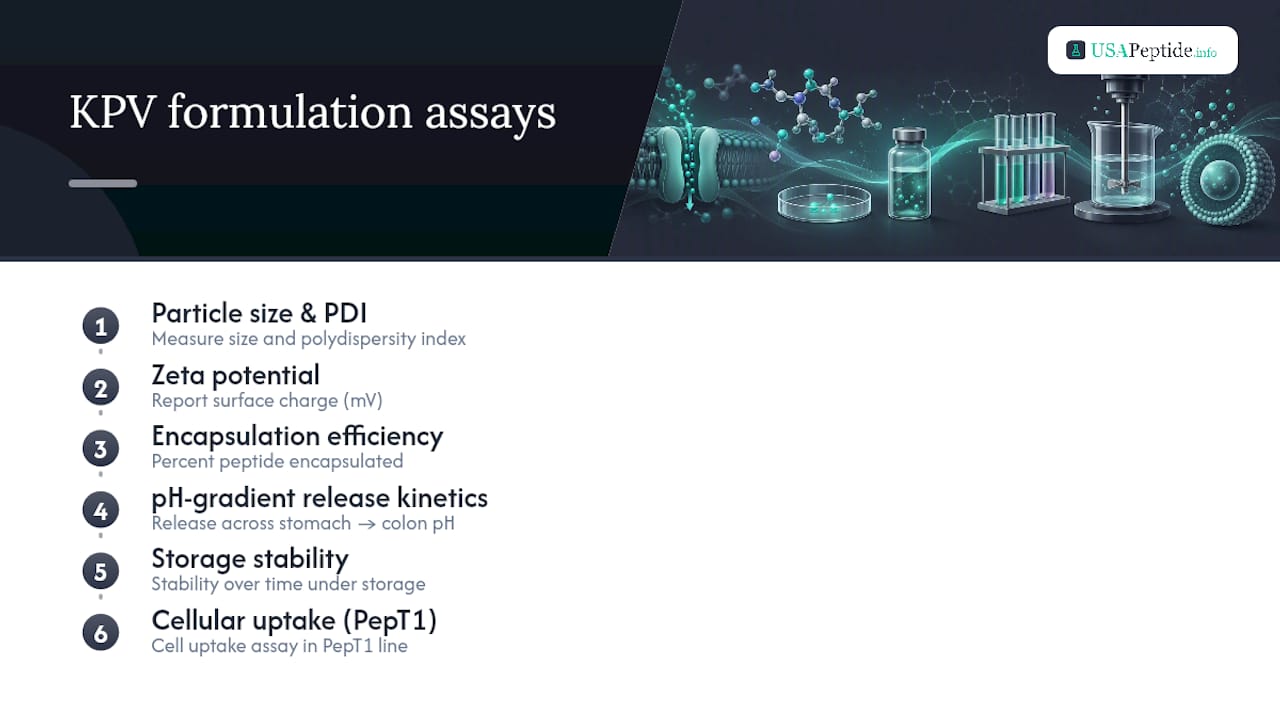
Anyone characterizing a new KPV formulation should report a consistent set of parameters: particle size and polydispersity index, zeta potential, encapsulation efficiency, in vitro release kinetics across a pH gradient mimicking stomach-to-colon transit, storage stability over time, and cellular uptake in a PepT1-expressing line. Without these, comparing one formulation's results to another's is close to meaningless.
Safety Data, Sourcing Risk, and Regulatory Status
No organ toxicity has been reported in the published mouse studies at the doses tested, and the peptide's lack of melanocortin receptor activity avoids the pigmentation and endocrine side effects associated with related α-MSH-derived compounds. That is a genuinely favorable preclinical safety signal. It is also the extent of what can honestly be said, because no human safety data exists. There are no completed dose-escalation studies, no pharmacokinetic profiling in human subjects, and no adverse event data collected under clinical trial conditions.
That evidence gap is precisely why KPV remains a research-use-only (RUO) compound. FDA guidance draws a firm line between materials intended for laboratory research and products approved for human therapeutic administration, and no synthetic peptide can cross that line without an approved pathway involving formal safety and efficacy review. Any product marketed for human consumption or clinical use falls outside that framework entirely.
Statistic Callout: The RUO peptide market carries a documented quality risk beyond regulatory status alone. Independent testing across peptide and adjacent compound categories has repeatedly found lot-to-lot purity variance and mislabeling, which means a certificate of analysis is not optional diligence. It is the only practical safeguard against running an experiment on a compound that isn't what the label claims.
Before procuring KPV for any experimental protocol, run through this sequence:
- Request the current Certificate of Analysis (COA) for the specific lot, not a generic product-page COA.
- Confirm HPLC purity is stated explicitly, ideally at or above 99%, and cross-check the retention time data if provided.
- Verify mass spectrometry data matches the expected molecular weight for Lys-Pro-Val.
- Confirm the supplier explicitly labels the product research-use-only, with no therapeutic or consumption claims anywhere in the listing.
- Store and handle according to the COA's stability recommendations, typically lyophilized and frozen until reconstitution.
Where KPV Research Needs to Go Next
The most obvious gap is also the hardest to close efficiently: nobody has run a controlled pharmacokinetic and pharmacodynamic study in a species larger than a mouse. Rat or non-human primate PK/PD data would clarify whether the 205 µg/day mouse dose scales in any predictable way, and it's a prerequisite before any first-in-human safety design becomes ethically defensible.
Second, human tissue work needs to move beyond the existing biopsy-level PepT1 expression data. Quantifying PepT1 density and activity across IBD severity grades in human colonic tissue would tell researchers whether the transporter-dependence finding from mouse knockouts actually predicts human responsiveness, or whether human PepT1 regulation under inflammation behaves differently.
Priority experiments worth funding, in rough order of translational value:
- Dose-ranging PK/PD studies in a second species to establish whether mouse dosing data scales linearly or requires allometric adjustment.
- Human colonic biopsy profiling of PepT1 expression across disease severity, correlated with existing IBD biomarker panels.
- A formal dose-escalation safety study design, even at the healthy-volunteer stage, incorporating standard tolerability and pharmacokinetic sampling.
- GMP-scale formulation reproducibility studies for the HA-PLGA nanoparticle system, since lab-scale synthesis rarely translates cleanly to manufacturing scale without re-optimization.
Pro Tip: Any study proposing to move KPV toward human testing should pre-register a biomarker panel, not just clinical outcome measures. MPO activity, histological inflammation scoring, and a standardized cytokine panel (TNF-α, IL-6, IL-1β at minimum) let you compare new data directly against the existing mouse literature instead of starting from zero.
Combinatorial designs also deserve more attention than they've gotten. Pairing KPV with an established mucosal-repair peptide, for instance, could test whether anti-inflammatory and tissue-repair mechanisms compound usefully, an approach comparable ongoing work on GI repair peptides is already exploring from the repair side.
Verifying Compound Quality for KPV Protocols
Every finding described above depends on knowing exactly what compound went into the assay. A COA with vague purity language or missing mass spec data undermines any conclusion drawn downstream, regardless of how careful the experimental design otherwise is.
USAPeptide's COA grading tool gives researchers a structured way to assess whether a supplier's Certificate of Analysis meets the standard needed for reproducible work, checking for complete HPLC purity reporting, mass spec confirmation, and lot-specific rather than generic documentation. The site's peptide dosage calculator supports experimental planning by scaling reference doses, like the 205 µg/day figure from the Dalmasso mouse study, to different body weights and administration routes for protocol design.
Beyond KPV-specific resources, a few tools support the broader research workflow:
- The peptide research glossary defines transporter and formulation terminology (PepT1, PLGA, zeta potential) for interdisciplinary teams working across pharmacology and materials science.
- ISO 17025-accredited testing and ≥99% HPLC purity standards, where a supplier states them explicitly, give researchers a verifiable baseline rather than a marketing claim.
- Cross-referencing a lot's COA against independent purity benchmarks before committing it to a multi-week colitis protocol avoids discovering a sourcing problem after the data is already compromised.
The Gap Between a Good Mechanism and a Proven Treatment
The PepT1 story is genuinely one of the more elegant pieces of peptide pharmacology in recent preclinical literature. A transporter that happens to be upregulated exactly where you want a drug to concentrate, carrying a peptide that hits NF-κB without touching melanocortin receptors, is not a common alignment of biology and convenience. We think that elegance is precisely why it gets oversold in less careful corners of the internet.
Mechanism is not efficacy, and mouse data is not human data. The honest position is that KPV deserves serious continued preclinical investment, particularly in larger-animal PK/PD work and human tissue profiling, before anyone frames it as anything beyond a research compound. Researchers moving in that direction should insist on verified analytical data at every step, because a contaminated or mislabeled lot doesn't just risk a bad experiment. It risks bad data entering the literature.
— USAPeptide Team
This article is general information, not a substitute for advice from a qualified doctor. Consult a qualified healthcare professional about your own circumstances before acting on anything here.
Sources
- PepT1-Mediated Tripeptide KPV Uptake Reduces Intestinal Inflammation
- FDA guidance documents (example resource)
FAQ
Does KPV Peptide Really Work?
In cell and mouse colitis models, yes. KPV reliably reduces inflammatory markers through PepT1-mediated uptake and NF-κB inhibition, but no completed human trials confirm this translates to people.
Who Should Avoid KPV Peptides?
Anyone outside a qualified research or laboratory setting should avoid KPV entirely, since it is sold research-use-only, lacks human safety data, and carries no approved clinical indication.
How Long Should You Take KPV Peptide?
There is no established human dosing duration because no human clinical protocol exists. Published mouse studies used continuous oral dosing (approximately 205 µg/day) over the course of a defined colitis induction period, not an ongoing regimen.
What Is the Best KPV Peptide Formulation for Research?
For colon-targeted preclinical work, hyaluronic acid-functionalized PLGA nanoparticles embedded in a chitosan/alginate hydrogel outperformed free KPV in mouse models by protecting the peptide and improving site-specific delivery.