BPC-157 promotes tissue repair primarily by driving angiogenesis through two coordinated pathways: VEGFR2-Akt-eNOS signaling at the cell surface and the recently identified FBXO22-BACH1 axis inside the nucleus. Supporting these core mechanisms are nitric oxide modulation, ERK1/2 and AKT pro-survival signaling, and measurable anti-inflammatory effects. Nearly all of this evidence comes from cell and animal models. Controlled human trials remain scarce, which matters for anyone trying to translate these findings into practice.
TL;DR:
- BPC-157's angiogenic effects primarily depend on VEGFR2-Akt-eNOS signaling and the FBXO22-BACH1 axis, which are influenced by dosage and administration route.
- Its molecular action involves increasing VEGFR2 expression, promoting vessel growth, and stabilizing BACH1 to enhance pro-angiogenic gene transcription.
- Short plasma half-life and route-specific metabolism mean mechanistic effects occur transiently and may involve active metabolites, requiring careful pharmacokinetic consideration.
- Safety risks stem from nitric oxide overproduction, potentially damaging mitochondria or affecting blood clotting, especially when combined with other NO-modulating therapies.
- Reagent quality verification, including purity and molecular identity checks, is essential for reliable mechanistic research on BPC-157.
Table of Contents
- What Is BPC-157, and Why Does Its Mechanism Matter?
- How Does BPC-157 Trigger Angiogenesis at the Molecular Level?
- Nitric Oxide, Antioxidant Enzymes, and the Double-Edged Sword of NO Signaling
- Cell Proliferation and Matrix Remodeling: The ERK, AKT, and FAK-Paxillin Connection
- What Anti-Inflammatory Effects Does BPC-157 Produce?
- How Is BPC-157 Metabolized, and Why Does That Affect Mechanistic Data?
- Preclinical Evidence vs. Human Data: Where the Gaps Are
- What Safety Risks Are Linked to BPC-157's Mechanism of Action?
- How Should Researchers Verify Reagent Quality Before Studying BPC-157?
- The Bigger Picture: Where BPC-157 Research Needs to Go Next
- Research Tools to Support Your BPC-157 Study Design
- Sources
What Is BPC-157, and Why Does Its Mechanism Matter?
BPC-157 is a synthetic pentadecapeptide, a chain of 15 amino acids derived from a protective protein found in human gastric juice. That origin explains a lot about its behavior in research settings: gastric-derived peptides tend to be unusually stable across a range of pH conditions, which is part of why BPC-157 shows biological activity even after oral exposure in animal models, an unusual trait for a peptide of its size.
Researchers studying BPC-157 mechanism questions run into a practical problem fast: the compound has been tested through several administration routes, and each route changes what you can actually conclude about its biological activity.
- Subcutaneous or intraperitoneal injection in rodent studies, the most common route in the orthopedic sports medicine literature, producing the highest and most consistent tissue concentrations.
- Oral gavage, used in gastrointestinal studies given the peptide's origin in gastric protective mechanisms.
- Topical or local application, occasionally used in wound-healing models to concentrate exposure at the injury site.
Why does this matter beyond academic interest? Because mechanism determines everything downstream in a research protocol. If a pathway like VEGFR2 activation only shows up reliably at certain tissue concentrations, then dosing schedules, sampling windows, and biomarker selection all need to be built around that pharmacokinetic reality rather than assumed from outcome-based studies alone. A researcher who treats BPC-157 as a black box that "heals things" will design weaker experiments than one who tracks the specific receptors and enzymes it engages. That distinction between outcome and mechanism is also why safety monitoring needs to be pathway-specific. Watching for generic "adverse events" tells you less than tracking the exact enzymes and receptors a compound is known to modulate. The peptide's full molecular profile, including sequence data and structural notes, is the starting point for designing that kind of targeted protocol.
How Does BPC-157 Trigger Angiogenesis at the Molecular Level?
Angiogenesis, the formation of new blood vessels from existing vasculature, sits at the center of BPC-157's regenerative profile, and it happens through two distinct but connected mechanisms.
The first is receptor-level activation. BPC-157 increases expression of VEGFR2 (vascular endothelial growth factor receptor 2) on endothelial cells and promotes its internalization, a step that triggers the downstream signaling cascade. Once VEGFR2 is activated, it phosphorylates AKT, which in turn activates endothelial nitric oxide synthase (eNOS). This VEGFR2-Akt-eNOS chain is the same core pathway that natural VEGF signaling uses to stimulate endothelial proliferation and tube formation, the structural first step of new capillary growth. Research published in the Journal of Molecular Medicine demonstrated this activation directly in endothelial cell assays, showing that BPC-157 exposure both upregulated VEGFR2 protein levels and drove functional tube formation in human umbilical vein endothelial cells (HUVECs), the standard cell model for angiogenesis research.
A 2026 study identified BPC-157 binding to FBXO22 at a specific residue (labeled P3), and showed this interaction blocks the normal ubiquitination process that would otherwise mark BACH1 for degradation. The result: BACH1 accumulates and drives transcription of pro-angiogenic growth factor genes, including PDGFB and FGFR1.
That second mechanism, the FBXO22-BACH1 axis, is the more recent discovery and arguably the more mechanistically interesting one. BACH1 is a transcription factor that normally gets tagged for destruction by the ubiquitin-proteasome system, with FBXO22 acting as part of that tagging machinery. Published in Cell Communication and Signaling, the study found that when BPC-157 occupies FBXO22, BACH1 escapes degradation and stays active in the nucleus, driving the transcription of genes that build new blood vessels. That's a meaningfully different kind of mechanism than receptor activation. Instead of amplifying a signal at the cell surface, BPC-157 is protecting a transcription factor from being destroyed, which then produces a sustained downstream transcriptional program rather than a short pulse of signaling.
Researchers confirmed the FBXO22 interaction with molecular docking simulations, surface plasmon resonance (SPR) binding assays, and alanine-scanning mutagenesis to map exactly which residues on BPC-157 make contact with FBXO22. That's a notably rigorous set of methods for a peptide mechanism paper. It moves the FBXO22-BACH1 finding well beyond correlation, into demonstrated direct binding. Complementary evidence came from chick chorioallantoic membrane (CAM) assays and hind-limb ischemia models, both of which showed increased vascular density following BPC-157 exposure, tying the molecular binding data to functional angiogenic outcomes in living tissue.

Nitric Oxide, Antioxidant Enzymes, and the Double-Edged Sword of NO Signaling
Nitric oxide (NO) sits downstream of the eNOS activation described above, and it does a lot of the actual physiological work: vasodilation, endothelial signaling, and cytoprotective effects that support tissue survival during repair. BPC-157's effect on NOS/eNOS expression has been documented across several preclinical models, with the narrative review in musculoskeletal healing research linking increased NO production directly to the angiogenic effects described in the previous section.
Alongside NO signaling, BPC-157 exposure correlates with induction of several antioxidant enzymes:
- Heme oxygenase-1 (HO-1), which breaks down heme and produces byproducts with anti-inflammatory and antioxidant properties.
- NAD(P)H quinone dehydrogenase 1 (NQO1), a detoxifying enzyme that protects cells from oxidative and electrophilic stress.
- Glutathione-related enzymes, part of the cell's primary antioxidant defense system.
Work summarized in PMC's mechanistic safety analysis ties HO-1 induction to reduced fibrosis in some tissue models, suggesting the antioxidant response isn't just protective housekeeping. It may actively shape how scar tissue forms during repair.
But NO signaling cuts both ways, and this is where mechanism-level thinking becomes a safety issue rather than just an academic one. Excess NO can react with superoxide to form peroxynitrite, a highly reactive molecule capable of damaging mitochondrial membranes and inhibiting components of the electron transport chain. Peroxynitrite formation has also been linked to disrupted heme and cytochrome P450 (CYP) enzyme function, which matters because CYP enzymes handle a large share of drug and hormone metabolism in the liver.
Pro Tip: If your protocol involves NO-modulating co-treatments alongside BPC-157, pair your primary endpoint with a nitrite/nitrate assay and a mitochondrial respiration readout. NO effects are dose- and context-dependent, and a single downstream marker won't tell you whether you're looking at cytoprotection or early oxidative stress.
Cell Proliferation and Matrix Remodeling: The ERK, AKT, and FAK-Paxillin Connection
Angiogenesis explains how new blood supply reaches injured tissue, but repair also requires the cells already at the site, fibroblasts, tenocytes, and myoblasts, to proliferate, migrate, and reorganize the surrounding matrix. BPC-157's mechanism here runs through a different but overlapping set of signaling nodes.
- ERK1/2 activation feeds into transcription factors including c-Fos, c-Jun, and Egr-1, all of which regulate genes controlling cell proliferation and early-response gene expression during wound healing.
- AKT signaling, the same node engaged downstream of VEGFR2, also supports cell survival independently of its angiogenic role, reducing apoptotic signaling in stressed cells at the injury site.
- FAK-paxillin complex involvement supports focal adhesion formation, the physical anchoring points cells use to migrate across the extracellular matrix during tissue remodeling.
Growth hormone receptor (GHR) upregulation adds another layer specific to musculoskeletal tissue. The systematic review of orthopedic sports medicine research documented increased GHR expression in tendon and ligament models following BPC-157 exposure, which may partly explain why so much of the animal literature on this peptide concentrates on tendon and ligament healing rather than generalized wound repair. Growth hormone signaling independently promotes collagen synthesis and fibroblast activity, so an upregulated receptor population would amplify whatever growth hormone is already circulating, rather than requiring BPC-157 to manufacture an entirely new signaling pathway from scratch.
Put together, ERK, AKT, and FAK-paxillin signaling form a proliferation and adhesion layer that runs in parallel to angiogenesis. New vessels bring blood supply; this second system determines whether the cells at the injury site can actually use it to rebuild functional tissue.
What Anti-Inflammatory Effects Does BPC-157 Produce?
Persistent inflammation is one of the main things that stalls tissue repair, so an anti-inflammatory mechanism matters just as much as a pro-angiogenic one. Across preclinical models, BPC-157 exposure correlates with measurable shifts in inflammatory biomarkers:
- Reduced cyclooxygenase-2 (COX-2) expression, the enzyme responsible for producing inflammatory prostaglandins at sites of tissue damage.
- Lower interleukin-6 (IL-6) and tumor necrosis factor-alpha (TNF-α) levels, two of the primary cytokines driving the acute inflammatory response.
- Decreased myeloperoxidase (MPO) activity, an enzyme marker of neutrophil infiltration and oxidative burst at injury sites.
The narrative review documents these reductions consistently enough across different injury models that the anti-inflammatory effect looks like a genuine mechanistic feature rather than an artifact of any single study design. Combined with the antioxidant enzyme induction covered earlier, HO-1 and glutathione-pathway activity, the overall picture is a tissue environment that's less inflamed and better buffered against oxidative stress during the repair window.
That combination has a plausible downstream effect on fibrosis. Chronic inflammation tends to drive excessive collagen deposition and scar tissue formation, so dampening the inflammatory signal earlier in the repair timeline could support more organized, functional tissue remodeling rather than dense scarring. This inference makes biological sense given the enzyme data, though it hasn't been directly confirmed through long-term functional outcome studies in humans.
How Is BPC-157 Metabolized, and Why Does That Affect Mechanistic Data?
Pharmacokinetics rarely gets the attention it deserves in peptide mechanism discussions, but it directly shapes how you should read every finding described above. BPC-157 undergoes hepatic metabolism with a short plasma half-life, according to pharmacokinetic data summarized in the orthopedic systematic review. The compound is cleared primarily through renal excretion, and mass spectrometry methods can detect it or its metabolites in animal tissue for a limited time following dosing.
That short half-life changes how mechanistic findings should be interpreted:
- A 30-minute plasma half-life means most of the signaling effects described in earlier sections, VEGFR2 activation, FBXO22 binding, ERK phosphorylation, likely occur during a narrow window after administration, not as a sustained steady-state exposure.
- Metabolic breakdown produces smaller peptide fragments and free amino acids, notably proline, which raises a real methodological question: are downstream effects attributable to intact BPC-157, its fragments, or the amino acids released during degradation?
- Route of administration changes tissue exposure patterns substantially. Oral dosing exposes gastrointestinal tissue directly but faces first-pass hepatic metabolism before reaching systemic circulation, while injection routes bypass that first pass but distribute differently across tissue compartments.
None of this undermines the mechanistic findings covered so far. It does mean that any researcher trying to correlate a specific pathway activation with a specific dose or route needs to account for a fast-clearing compound with metabolites that may carry independent biological activity of their own.
Preclinical Evidence vs. Human Data: Where the Gaps Are
The mechanistic case for BPC-157 is dense on the preclinical side. Angiogenesis assays, inflammatory biomarker panels, receptor-binding studies, and now direct transcription factor interaction data all point in a consistent direction across independent labs and injury models. Controlled human trials testing these same mechanisms are, by comparison, essentially absent from the peer-reviewed literature.
That gap creates specific translational problems that go beyond the usual "more research is needed" caveat, as discussed in detailed commentary on peptide research trends that highlights practical translational considerations.
- Dosing translation is unresolved. Effective doses in rodent models don't automatically scale to human dosing through simple weight-based conversion, particularly for a peptide with route-dependent bioavailability.
- Metabolic variability between species matters. Hepatic metabolism rates and half-life data from rodent studies may not transfer directly to human hepatic clearance.
- Commercial peptide preparations vary widely in quality. A mechanistic finding from a controlled academic lab using a verified reagent doesn't tell you anything about a peptide product manufactured under different quality standards.
- No standardized biomarker panel exists for human trials. Without agreed-upon endpoints (VEGFR2 phosphorylation levels, NO metabolite panels, inflammatory cytokine measurements), it's difficult to compare findings across the handful of small-scale investigations that do exist.
Investigators pushing this field toward clinical relevance have specific design elements they need to prioritize. Robust pharmacokinetic and pharmacodynamic (PK/PD) modeling in early-phase human studies would establish actual half-life, metabolite profiles, and dose-response relationships, rather than assuming rodent data transfers directly. Trials also need pre-specified biomarker panels tracking the exact mechanisms described throughout this article, not just subjective outcome measures like pain scores. Safety endpoints need to specifically monitor NO-related and cardiovascular markers given the mechanistic risks discussed below, not just generic adverse event reporting. As one analysis on mechanistic discovery framed it, the field is shifting from broad observation of regenerative outcomes toward identifying precise intracellular targets, and that shift is exactly what makes rigorous, biomarker-driven human trials both more feasible and more necessary.
What Safety Risks Are Linked to BPC-157's Mechanism of Action?
The same NO-centered mechanism that explains BPC-157's angiogenic and cytoprotective effects also defines its main theoretical risk profile. This isn't a separate safety conversation bolted onto the mechanism data. It's a direct extension of it.
- Mitochondrial and oxidative risk. As covered earlier, excess NO can generate peroxynitrite, which damages mitochondrial membranes and can interfere with heme and CYP enzyme function, raising theoretical concerns in contexts of extreme or dysregulated NO signaling.
- Cardiovascular considerations. NO is a potent vasodilator, and eNOS-driven NO production affects platelet aggregation. Anyone combining BPC-157 with other agents that modify eNOS activity or blood clotting should treat that as a mechanistically plausible interaction, not a hypothetical one.
- CNS signaling uncertainty. Preclinical work has linked BPC-157 to modulation of dopaminergic and serotonergic neurotransmission, a finding worth tracking but far from established in human contexts.
Pro Tip: Build your safety monitoring panel around the mechanism, not just general toxicity screening. Liver function tests make sense given hepatic metabolism, oxidative stress markers (nitrite/nitrate, malondialdehyde) track the NO pathway directly, and basic cardiac and hematology panels catch vasodilation or platelet-related effects early. Also run purity verification on your peptide source itself, since contaminants can produce confounding results that look like a drug effect but aren't.
How Should Researchers Verify Reagent Quality Before Studying BPC-157?
Mechanistic conclusions are only as reliable as the reagent that produced them. A peptide sample with unverified identity or contamination can generate signaling data that looks meaningful but reflects impurities rather than BPC-157 itself, a concern echoed in guidance on peptide stability and reagent verification.
- Check HPLC purity data on the Certificate of Analysis. Research-grade peptide work generally expects purity at or above 99%, verified by high-performance liquid chromatography, not just a manufacturer's stated concentration.
- Confirm identity verification methods. Mass spectrometry confirmation of molecular weight, alongside HPLC, gives you two independent lines of evidence that the vial contains what the label claims.
- Watch for inconsistent lot data or missing test dates, both common red flags for suppliers cutting corners on quality control.
- Build in the right positive controls for your mechanism of interest. VEGFR2 phosphorylation assays, eNOS activity measurements, BACH1 protein level tracking via western blot, and ROS assays all give you direct, mechanism-specific readouts rather than relying on downstream phenotypic outcomes alone.
USAPeptide's BPC-157 reference page includes molecular data alongside a COA grading tool built specifically to help researchers assess whether a Certificate of Analysis meets these identity and purity standards before it becomes the basis for a mechanistic study.
The Bigger Picture: Where BPC-157 Research Needs to Go Next
The molecular case for BPC-157 has gotten considerably stronger with the FBXO22-BACH1 discovery. It gives researchers a specific, testable transcriptional target rather than a vague "pro-healing" label, and that specificity is genuinely useful. But strong preclinical mechanism data is not the same as clinical validation, and conflating the two is where a lot of the public conversation around this peptide goes wrong.
We think the field's priority now should be biomarker-driven early-phase human studies that track the exact pathways described here, VEGFR2 phosphorylation, NO metabolites, BACH1 stabilization, rather than another round of animal studies confirming what's already reasonably well established. Multicenter replication and standardized reporting on reagent purity would also do more for this field's credibility than another single-lab rodent study. Until that work exists, treat BPC-157's mechanism as promising and well-characterized at the molecular level, not as a settled clinical fact.
— USAPeptide Team
Research Tools to Support Your BPC-157 Study Design
If you're designing a study around any of the pathways covered here, VEGFR2 signaling, NO modulation, ERK/AKT proliferation markers, the practical bottleneck is usually reagent quality and dosing precision, not the mechanism itself. USAPeptide.info exists specifically to close that gap for researchers rather than sending you to scattered manufacturer claims with no independent verification.
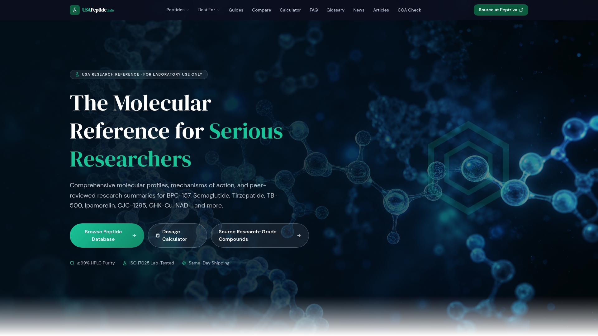
The BPC-157 molecular profile page lays out sequence data, mechanism summaries, and links to quality documentation in one place, rather than requiring you to cross-reference five different sources. The COA grading tool lets you check a Certificate of Analysis against expected purity thresholds and identity markers before you commit a reagent to a study protocol. If your research spans multiple peptides related to tissue repair, the recovery-focused peptide guide maps out mechanistic overlaps and differences across compounds, useful context when you're deciding which peptide fits a specific pathway you're investigating. None of these tools replace your own experimental controls or a clinical judgment call. They're built to support rigorous research design, not to make treatment recommendations. Start by pulling up the BPC-157 reference page and running your current supplier's COA through the grading tool before your next batch order.
Sources
The mechanistic claims in this article draw primarily on peer-reviewed molecular and cell-based research. For a deeper look at the VEGFR2 activation data, see the original study in the Journal of Molecular Medicine. The FBXO22-BACH1 discovery is documented in full in Cell Communication and Signaling. For broader context on preclinical evidence and translational limits, the systematic review in orthopedic sports medicine and the narrative review on musculoskeletal healing both offer detailed evidence synthesis, alongside additional mechanistic and safety discussion in PMC's analysis of NO and antioxidant pathways.
This article is general information, not a substitute for advice from a qualified doctor. Consult a qualified healthcare professional about your own circumstances before acting on anything here.
- Therapeutic potential of pro-angiogenic BPC157 is associated with VEGFR2 activation and up-regulation | Journal of Molecular Medicine
- Emerging Use of BPC-157 in Orthopaedic Sports Medicine: A Systematic Review
- BPC157 drives angiogenesis through FBXO22-dependent stabilization of BACH1 | Cell Communication and Signaling